
Growing Up
Award: Semifinalist, Images of Research Competition 2018
| Submitted by: | Gregory Funston |
| Faculty/Department: | Faculty of Science, Department of Biological Sciences |
| Place of creation: | Dino Lab, University of Alberta |
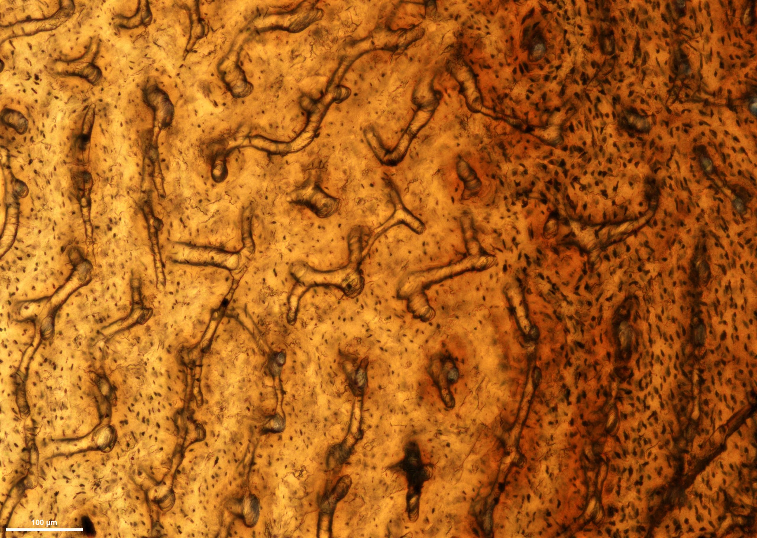
This image is a thin-section of a fossilized dinosaur shin bone. The bone belongs to a feathered theropod dinosaur called a caenagnathid, which would have looked like a colourful ostrich or emu with a long tail and a crest on its head. Thin-sections are one of the best ways of understanding the physiology of extinct animals. As bones grow, they leave behind telltale marks that allow us to understand the growth rate and age of the animal. In addition to the large tunnels in the images, which are vascular canals for blood vessels that fed the bone, you may notice hundreds of small black dots. These black dots are called osteocyte lacunae, and they record the spaces filled by bone-secreting cells. The change in their size and density towards the darker righthand side of the image represents some physiological change within the first year of this individual’s life. While there is not enough evidence to be certain, it likely represents fledging, when the animal left the nest and stopped receiving parental care. Unfortunately, life must have been hard for this little creature on its own: it died and was fossilized shortly after its first birthday.