
Semi-finalist 2025
| Submitted by: | Allison Thome |
| Department: | Chemistry |
| Faculty: | Science |
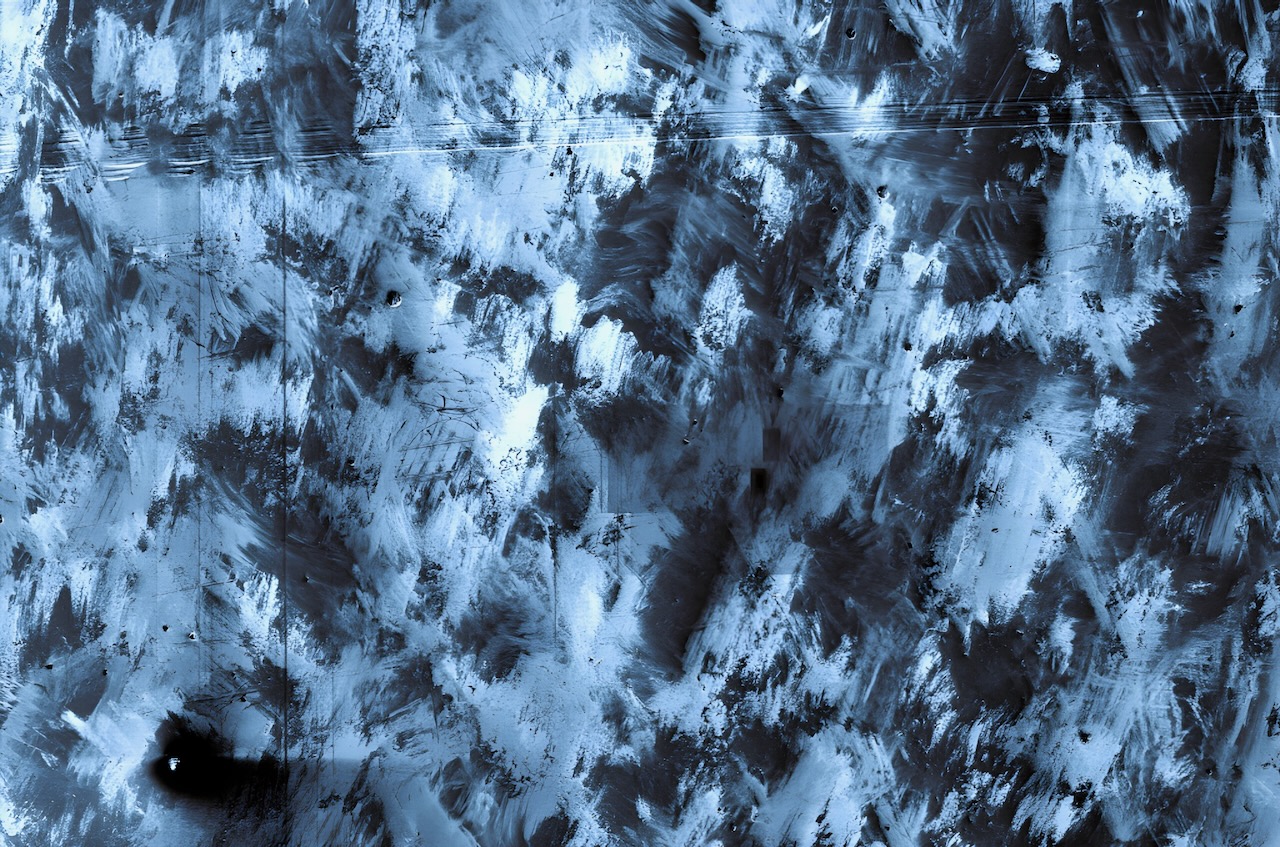
The image reveals the secret artistic life of a laser printed stainless steel sample caught in its “un-presentable” state, before it has been thoroughly cleaned. It highlights a hidden world of abstract expressionism that exists on every sample but is often unseen. The dynamic swirls, scratches and textures are created by polishing compounds, grease, and other contaminants that form a composition across the steel canvas. Though striking, this art needs to be erased to give way to another critical imagery beneath: the grain structures that tell the true story of the material’s printing process. The accidental artistry has to be removed before the deliberate scientific analysis can commence.
My research characterizes these advanced manufactured materials, requiring spotless surfaces for accurate analysis. Yet there’s a contradiction knowing that before every publishable microscopy image exists this temporary gallery of accidental art. This image celebrates that paradox – the moment when scientific preparation and art intersect, reminding us that scientific process contains hidden moments of unexpected beauty typically lost in the pursuit of publishable data.
Was your image created using Generative AI?
No.
How was your image created?
This image was captured using a ThermoFisher Helios Hydra PFIB/SEM (Plasma Focused Ion Beam/Scanning Electron Microscope) at the nanoFAB Characterization Centre. The sample, a laser-directed energy deposition printed stainless steel cubic specimen, was sectioned and coarsely polished using standard metallographic preparation techniques.
The microscopy was performed at 200x magnification using an accelerating voltage of 20.00 kV and a beam current of 0.40 nA. The High-performance in-chamber electron and ion detector (ICE) was employed to collect the signal, which is sensitive to both topographical and compositional differences. The color contrast was adjusted and the grayscale SEM image was digitally colorized with a blue tint gradient using Microsoft Photos application, applying a monochromatic color overlay to transform the standard black and white electron microscopy data into a more visually striking representation. This color treatment was deliberately chosen to emphasize the textural patterns and enhance the abstract expressionist quality while preserving all original structural information captured by the electron beam microscope.
Where is the image located?
ThermoFisher Helios Hydra PFIB/SEM Microscopy Suite, nanoFAB Characterization Centre, University of Alberta. The image was collected during surface characterization analysis of laser-directed energy deposition printed stainless steel samples as part of materials science research.