
Submission 2023
| Submitted by: | Dale Archer |
| Department: | Biological Sciences |
| Faculty: | Science |
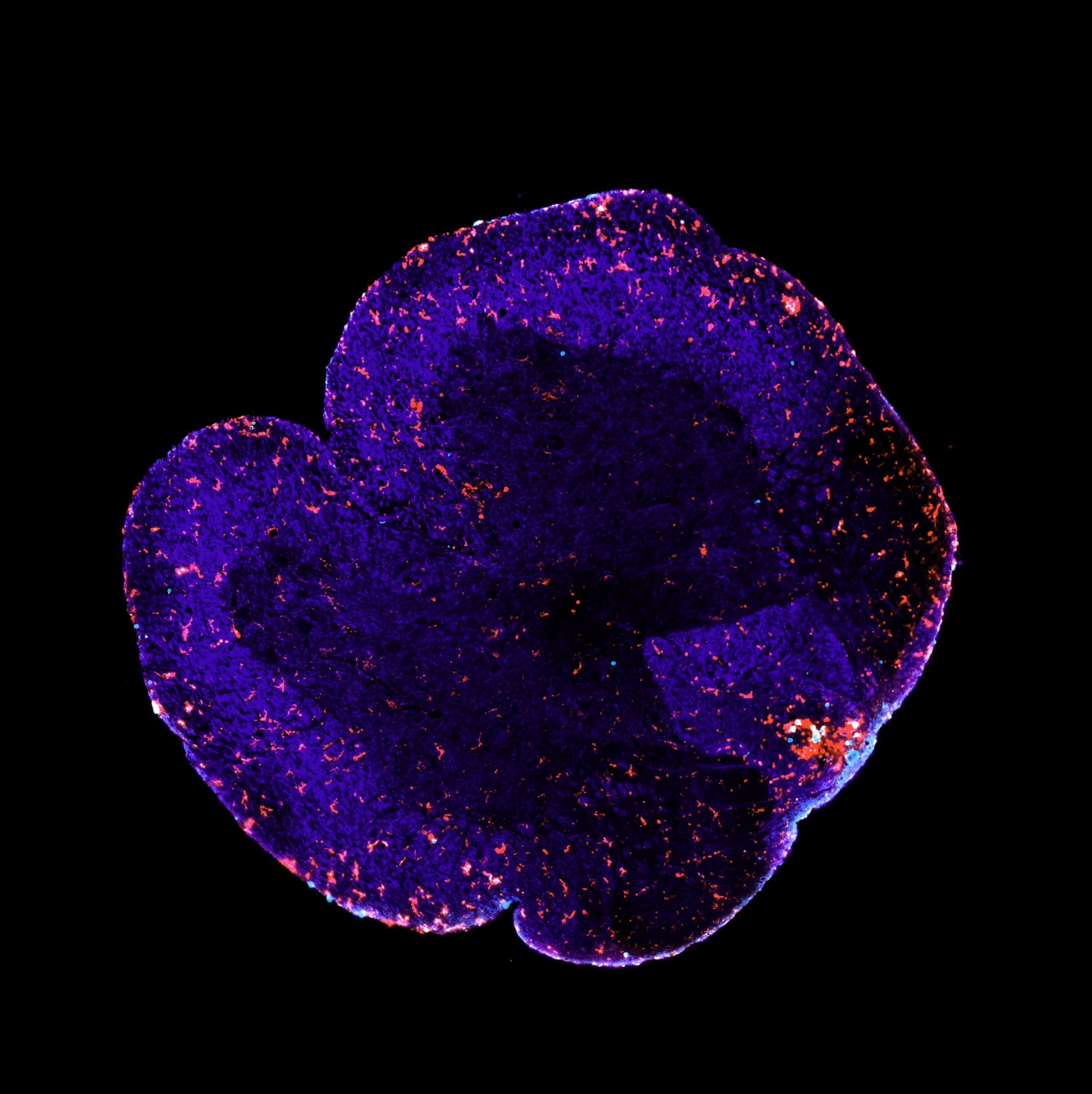
This is a section of a spinal cord from a mouse that experienced similar pathology and symptoms as a multiple sclerosis (MS) patient. Multiple sclerosis is an autoimmune disease characterized by a loss of myelin (demyelination) in the brain and spinal cord. Using immunofluorescent staining we can visualize the myelin (in dark blue) as well as immune cells that drive inflammation and demyelination, including microglia and macrophages (in red) and T cells (in light blue). The aggregation of microglia/macrophages and T cells and the faint myelin staining seen in the bottom right of the image is an inflammatory lesion where there is active inflammation and demyelination. In our research, we are interested in determining the effects of gut microbes that are not commonly found in industrialized people (termed VANISH species) on disease pathology in a mouse model of MS. Immunofluorescent staining and imaging allows us to assess the ability for these microbes to modulate disease pathology in these mice. The findings from this research will be informative for future development of microbial-based therapeutics targeted for MS patients.