Submission 2025
| Submitted by: | Aria Khalili |
| Department: | Mechanical Engineering |
| Faculty: | Engineering |
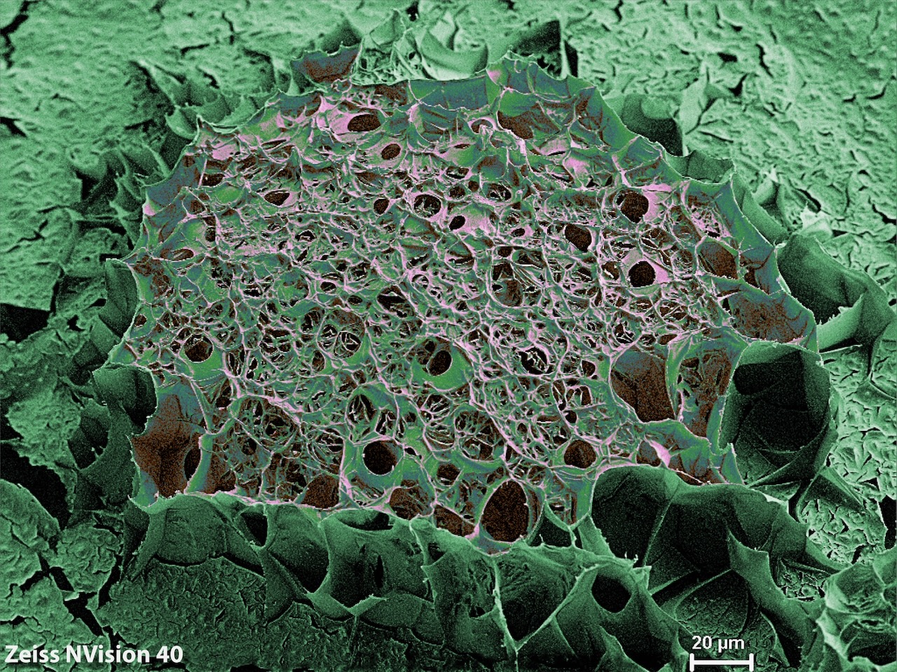
This cryo-SEM image reveals the hidden architecture of a lignin-gelatin hydrogel bead, captured to advance imaging methods for soft material research. Through rapid freezing in liquid nitrogen, its delicate structure was preserved, exposing a web of interconnected pores that weave across the surface. I manually enhanced the image with color to emphasize these intricate features, merging scientific precision with visual storytelling. This bead serves as a model for exploring how cryo-SEM can unlock the fine details of soft, hydrated materials—structures that often elude conventional imaging. The work bridges materials science with visualization, helping to refine techniques for studying complex biological and synthetic hydrogels.
Was your image created using Generative AI?
No.
How was your image created?
The image was captured using a cryogenic scanning electron microscope (cryo-SEM). A lignin-gelatin hydrogel bead was plunge-frozen in liquid nitrogen to preserve its native structure. The sample was sputter-coated with a thin layer of conductive metal (Pd/Pt) using a Leica EM ACE600 coater to prevent charging. Imaging was performed in secondary electron mode at 3 kV accelerating voltage and 2 mm working distance. The sample was kept below −130 °C during imaging to maintain structural integrity. After acquisition, the grayscale SEM image was manually color-enhanced using image editing software to highlight pore structures and enhance visual clarity.
Where is the image located?
National Research Council of Canada, Edmonton, Alberta, during microscopic characterization of hydrogels.