Semi-finalist 2025
| Submitted by: | Henry Sharpe |
| Department: | Biological Sciences |
| Faculty: | Science |
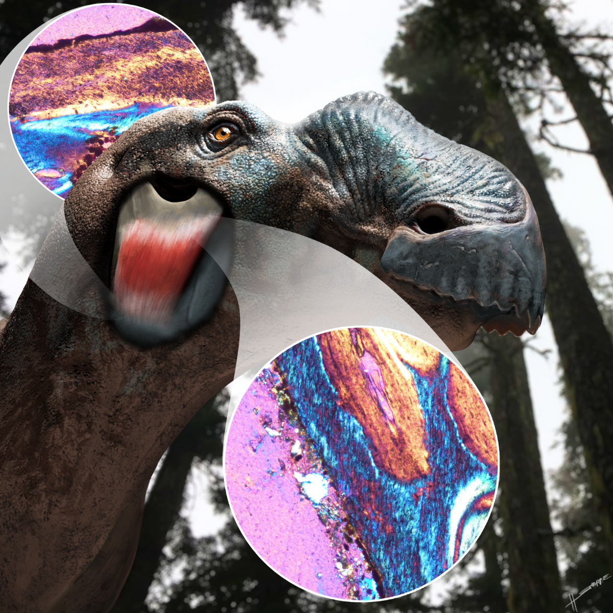
Dinosaur soft tissues usually don’t preserve in fossils, so paleontologists must use nuanced methods to reconstruct them. Muscles and ligaments will insert collagen fibres into bone to anchor themselves, and these fibres can remain in the bone after death and fossilization. Sharpe et al. (2025) histologically sectioned dinosaur cheek and jaw bones to find these fibres, and then traced their angle of entry in 3D. Complementary fibres in the cheekbones and mandible (the scratchy texture beneath the bone surface in the histological slides) revealed a previously-unknown muscle or ligament in the cheek of almost all dinosaurs, termed the exoparia. This image displays a duck-billed dinosaur from the Cretaceous of Alberta, showing the exoparia in its anatomical position, and two of the histological slides that revealed its presence after 75 million years.
Was your image created using Generative AI?
No.
How was your image created?
The background image is a photograph taken (on personal time) on Vancouver Island, British Columbia, which has a superficially similar flora and environment to Cretaceous Alberta. The dinosaur was created by first surface scanning a skull (housed in the Canadian Museum of Nature, Ottawa), and then sculpting soft tissues in Autodesk Maya and ZBrush, and finally painting colours and texture details in Adobe Photoshop. The histological slides were created by thin-sectioning dinosaur bones from the Royal Tyrrell Museum and University of Alberta collections, and they were photographed under cross-polarized light using a compound microscope, a Nikon microscope-mounted camera, and NIS-Elements D. All elements (background photo, dinosaur, histological slides) were then combined and composited in Adobe Photoshop, and transparent solids were added to indicate the position of the histologically-sectioned bones in vivo.
Where is the image located?
A visualization of a dinosaur soft tissue created by photography (Vancouver Island, British Columbia), 3D modelling (Sullivan Lab, U of A), digital painting (Caldwell Lab, U of A), and histology (Vert.Paleo. histology lab, U o fA) of bones from Dinosaur Provincial Park, Alberta.