Submission 2023
| Submitted by: | Elmira Khiabani |
| Department: | Psychology |
| Faculty: | Science |
| Collaborator: | Anna Kalisvaart |
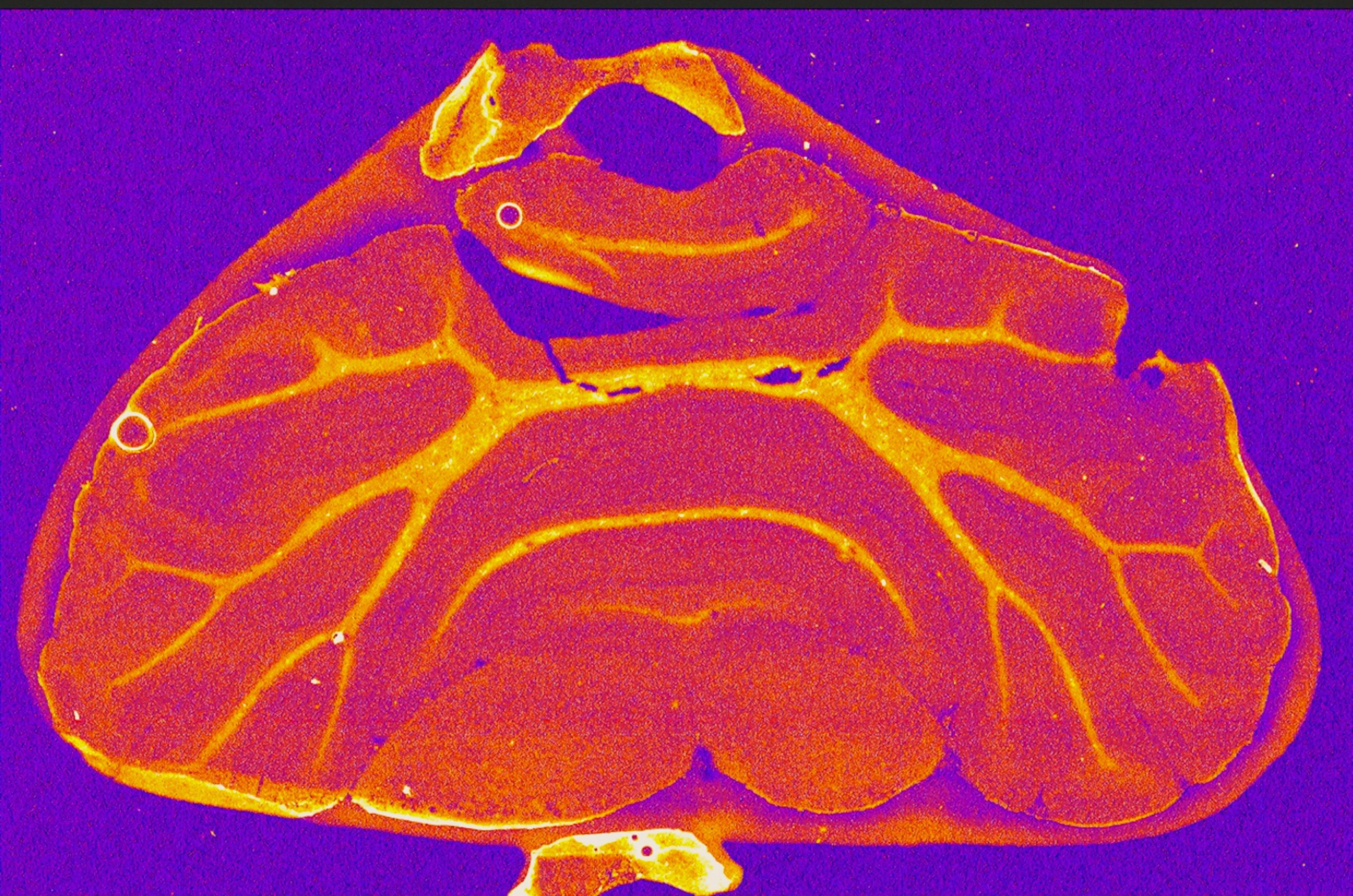
This is an image of the rodent cerebellum captured with X-Ray Fluorescence Microscopy, a method that produces beams of light a million times brighter than the sun. This beam excites electrons within a sample to emit photons to generate elemental maps, such as this one.
The brain is the most complex organ of our body and a vital source of human function, controlling everything from thoughts and emotions to physical movements. The complexity of the brain is generated by the collective effort of individual cellular building blocks. Cells require tight regulation of their ionic environment; however, this careful balance is disrupted by brain injuries, such as stroke, resulting in dysfunctional neural activity. As a stroke researcher, I use advanced imaging techniques to examine disturbances in ionic homeostasis, and how this drives cellular abnormalities in damaged brain regions.
Microscopy reveals an often hidden, yet beautiful side to science, allowing us to appreciate how the smallest cellular units of life serve a vast role in dictating human function and experience. As we have here, we can image specific brain regions to observe a vibrant rainbow of colour that is representative of its elemental make up. Generating images like these are a crucial step in understanding how to improve outcomes and formulate treatments for those affected by stroke and other brain injuries.