
Submission 2023
| Submitted by: | Zhe Lyu |
| Department: | Chemical and Materials Engineering |
| Faculty: | Engineering |
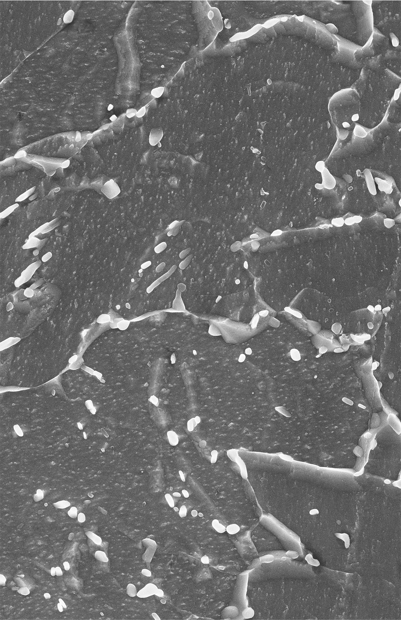
This image, taken by a scanning electron microscope (magnified × 10k), shows the microstructure of a service-exposed low alloy steel exposed to a high temperature and pressure environment. The distinct contrast between the grey ferrite matrix and the white particles in the image allows for detailed analysis of the microstructural features, including the morphology and spatial arrangement of the carbides. These carides are developed during service, and this image serves as a visual representation of the microstructure, aiding in the understanding of the material’s mechanical properties, and contributing to the ongoing research efforts in the field of metallurgy.