Semi-finalist 2023
| Submitted by: | Matthew Martell |
| Department: | Electrical and Computer Engineering |
| Faculty: | Engineering |
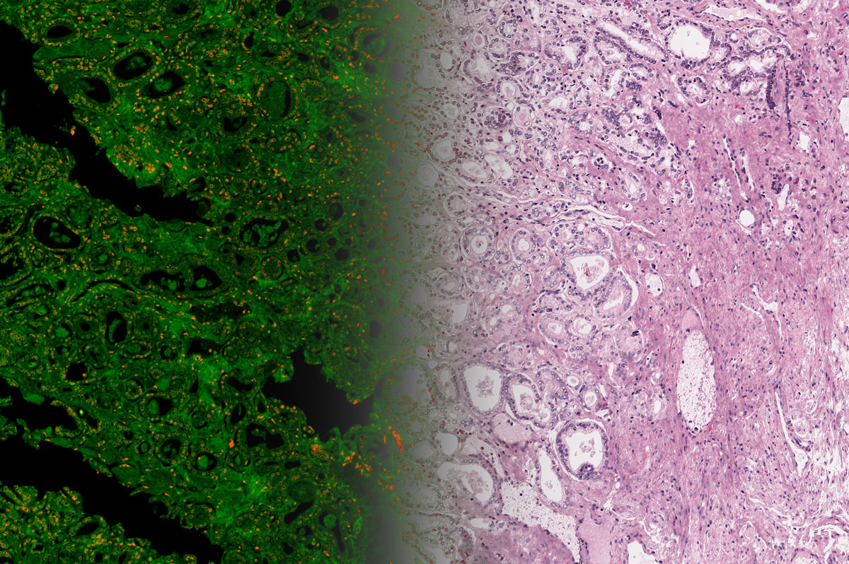
For over a century, pathologists have relied on a technique called H&E staining, the gold standard for cancer diagnosis. Unfortunately, this time-consuming process often requires days to chemically dye important tissue features in different colours for visualization under a microscope. This means it cannot offer guidance during surgery to ensure complete tumor removal, and many patients currently face the trauma and risk of follow-up surgeries when residual cancer is found post-operatively. To address this problem, my research aims to develop a new microscope that bypasses the staining process using ultraviolet laser light. As shown in this prostate tissue sample, our method generates images (left) of intrinsic UV light absorption in orange and reflection in green, which correspond well to features in a true H&E stain. We then use machine learning to transform and render this information in a style (right) nearly indistinguishable from the true H&E stain familiar to pathologists for interpretation. This stain-free virtual H&E imaging has the potential to provide rapid feedback to surgeons, verifying complete tumor removal during the first procedure to improve outcomes for patients.