
Submission 2024
| Submitted by: | Soroush Mirkiani |
| Department: | Neuroscience |
| Faculty: | Medicine & Dentistry |
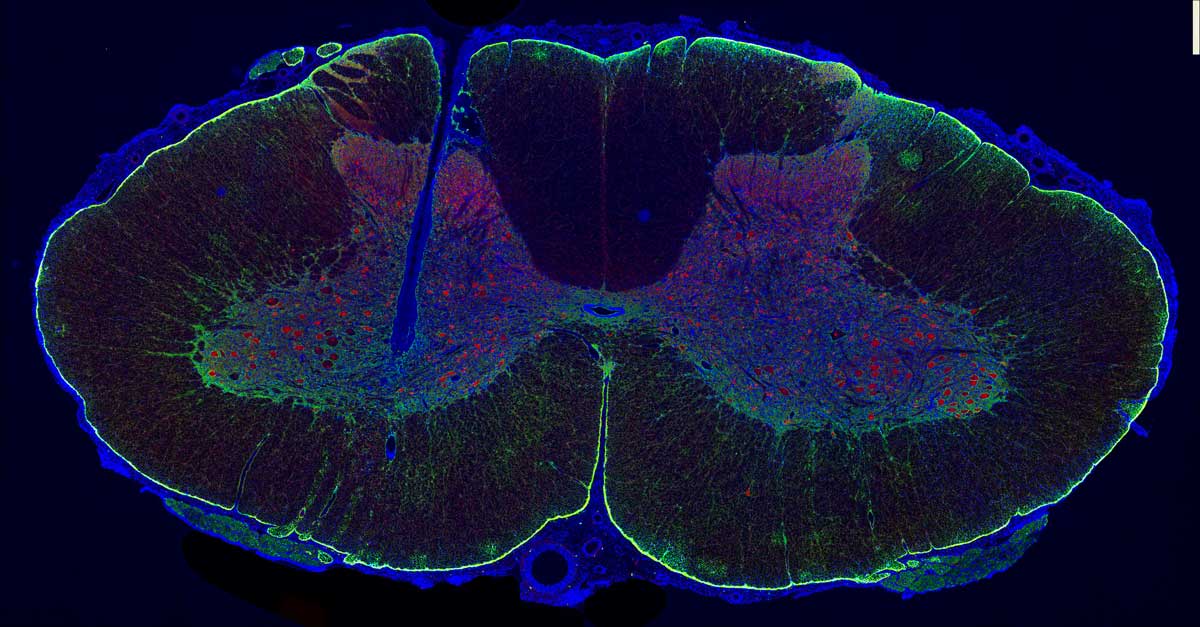
Under the microscope’s eye, a spinal cord section resembles a bird’s-eye view of a city. Neurons’ red glow mirrors scattered home lights, each a node in spinal cord activity’s network. Astrocytes, in green, support neurons like roads or infrastructures between homes, ensuring efficient function within the spinal cord network. Microglia, stand as vigilant guardians in the spinal cord, always watchful for potential threats or disturbances. To the left, a dark valley cuts through the city. Microglia, with their blue nuclei, encircle it like crime scene tape. However, appearances can be deceiving, and not everything is as it seems. After a spinal cord injury, the connection between brain and body is disrupted. Yet, rays of hope shines again on the city of spinal cord. Electrical stimulation below the injury level could awaken muscles, and restore standing and walking ability. While microelectrodes insertion may cause minor injuries, they are actually helpful. Glial cell activity ensures that while some may fall, the city remains resilient in its desire for restoration.
Captured from domestic pig spinal cord tissue after 8 days of microelectrode implantation.
Was your image created using Generative AI?
No.
How was your image created?
Epifluorescence microscopy was used for capturing this image. First, fluorescent dyes or proteins are used to label cellular components in the frozen and sectioned tissue slices. Epifluorescence microscopy enhances immunohistochemistry by detecting the fluorescence emitted by antibodies. It works by illuminating samples with excitation light, exciting fluorescent molecules that emit light at longer wavelengths. Fluorescent signals are captured by a detector, producing images. Epifluorescence microscopes have various filters that offers higher contrast imaging, enabling selective visualization of labeled structures against a dark background. It supports simultaneous visualization of multiple labels. We can also quantitatively analyze fluorescence intensity and distribution. Epifluorescence microscopy is a tool for studying cellular localization and protein expression in biological samples, aiding research in various fields such as neuroscience, cancer biology, and immunology.