
Submission 2024
| Submitted by: | Masha Masha |
| Department: | Cell Biology |
| Faculty: | Medicine & Dentistry |
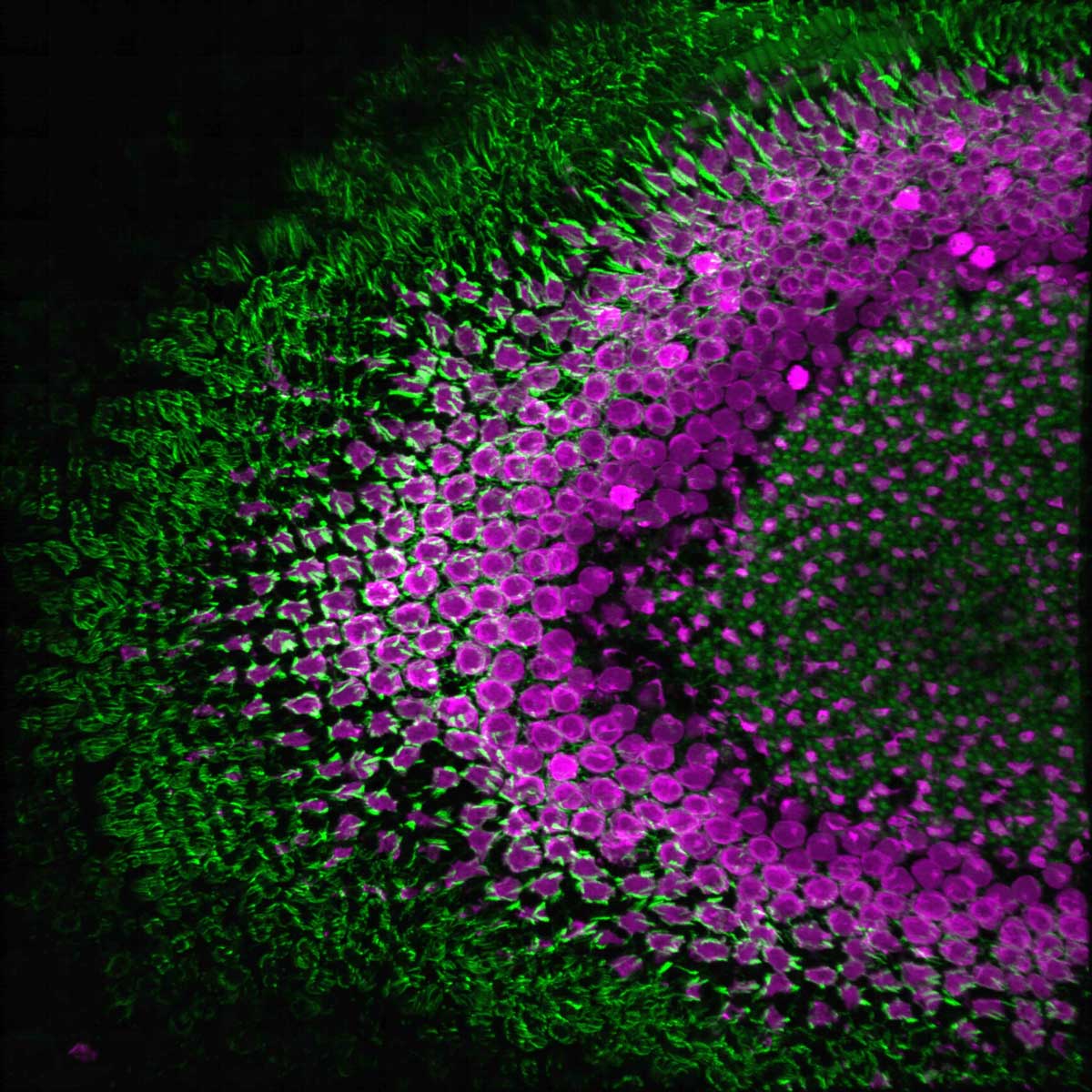
My research focuses on finger-like protrusions around the sensory ending of photoreceptors, the primary light-sensing cells of the retina. As those protrusions have a core of common structural protein called actin, I usually visualize them by staining the tissue with the actin-labeling dyes. This image depicts a transverse section through a transgenic Tg(sws1:GFP) zebrafish retina (UV-sensitive cone photoreceptors in magenta), demonstrating the distribution of phalloidin-stained actin (green) in the photoreceptor layer. 3D reconstruction of a confocal stack.
Was your image created using Generative AI?
No.
How was your image created?
Zebrafish at 1 month post-fertilization were euthanized and fixed in paraformaldehyde, then washed and prepared for freezing using sucrose. The fish were then placed into molds and frozen in a special medium. Afterwards, I cut the block on a cryostat and collected sections on a slide. I performed a standard immunohistochemistry protocol on those slides and mounted them. Later, I imaged the slides on a confocal microscope and used Imaris software to create a 3D reconstruction.