
| Submitted by: | Amir Adel |
| Department: | Biomedical Engineering |
| Faculty: | Medicine & Dentistry |
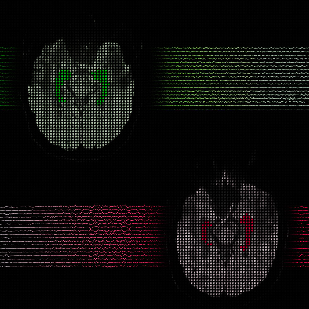
Do you see the coloured pairs that look like seahorses? Imagine looking from the top at horizontal cuts in the human brain. When the anatomist, Arantius, saw the brain of a cadaver, he named each of those regions hippocampus or “seahorse” in Greek. Here, you are looking at brain images of two living individuals obtained by magnetic resonance imaging (MRI). The way your hippocampi appear in MRI gives physicians clues about your health.
Epilepsy is a chronic disorder, with its hallmark of recurrent seizures. Doctors detect a seizure using recordings of brain electrical activity – those wiggly lines. Unfortunately, many epilepsy patients undergo an invasive brain surgery that doesn’t help much. What if we had a tool that enabled better selection of patients for epilepsy surgery? My research uses an advanced MRI method to answer this question. I study the architecture of the hippocampus in epilepsy patients since the seahorse is a key determinant of surgery success in these patients. Ultimately, advanced MRI may allow doctors and patients to make informed decisions about whether surgery is the right choice.
To a brain imager’s eye, each pixel is significant. Zoom in, what do you see?