
Submission 2025
| Submitted by: | Jennie Vu |
| Department: | Physiology |
| Faculty: | Medicine + Dentistry |
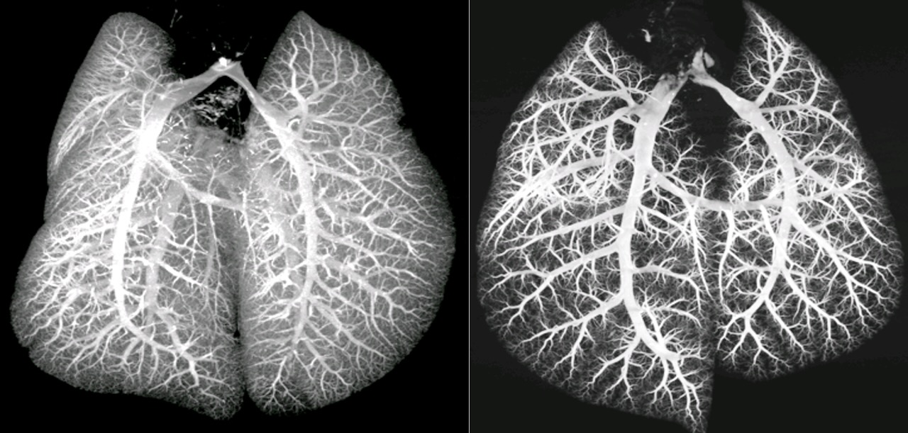
This image depicts lung vasculature of a healthy control rat (left) in contrast to the lung vascular bed of a diseased rat with pulmonary arterial hypertension (right). My project investigates how lung vasculature adversely remodels in pulmonary arterial hypertension. Pulmonary arterial hypertension resulted in the loss of vessel luminal volume and branching, which means there is an overall diminution of vessel volume that is perfused with blood along with reduced vessel arborization.
Was your image created using Generative AI?
No.
How was your image created?
Rat lungs were perfused with a barium contrast-gelatin suspension and then fixed with paraformaldehyde. Perfused lungs were then visualized with micro-computerized tomography angiography which images the lungs in high resolution thin sections and computationally generates a 3D rendering of the perfused vessels of the lungs using a pi-mod reconstruction and analysis software.
Where is the image located?
This image was taken in a micro-computerized tomography machine by MILabs located in the Katz basement.